Showing posts with label Period 7. Show all posts

Friday, April 1, 2011

Pea Soup!


First Mr. Finley told us we had to write a paper connecting genetics with meiosos and mitosis that has 12 font, double spaced, and is 1 page. It has to be double spaced. We then started talking about the Pea Soup! experiment. We didn't do a great job identifying patterns and similarites and differences we have already seen. Some were that all the green peas had yy letters and the yellows had Yy and YY. The yellow peas have atleast 1 uppercase Y. All the round peas had atleast 1 uppercase R. This means its round. Evidence its only 1 is the parent has 2 RR and the children are rR and they are all round. We chose to have a yellow pea thats round by itself. The children of it should have 3 yellow peas and 1 green pea. 3 will be round, and 1 will be wrinkled. We ended up with 2 yellow round, 1 yellow wrinkled, and 1 green round. We then discussed how to be able to predict transmission. Since 50% of chromosomes come from the parent and 50% come from the other parent. The method to figure out colors are that the first child gets the first Y's from the first ys of the parents. The second child gets the first Y from the first parent and second from the 2nd parent. The 3rd child gets its first letter from the 1st parents 2nd letter and 1st letter from the 2nd. The last child gets the 2nd letters from the parents. We figured out that the letters represebt chromosomes. To explain that 50% chromosomes come from each parent is that the egg and sperm are haploids. So you end up with 2 sperm with the first letter, 2 with the second. And the other 2 eggs with the first letter, 2 with the second.
IW

Tuesday, March 8, 2011

3/8/11 stations


Today we started class by taking out our lab sheets from yesterday. Mr. finley said then that we were going to be doing station work today. Our group first had to do station 1 which was completing questions in the textbook on page 97. Another group was at the smartboard with Mr. Finley, and the last group was online at cellsalive.com on the mitosis site. At station 1 the group went over some qustions about mitosis based on a graph with the amount of time the cell spends in each section. Some of the questions were like how long is the cell in interphase and how long is mitosis. Our group got done with all the multiple choice questions and then got to some open-ended ones about how to set up an experiment to test for mitosis while changing a ariable. We said that you could do one plant under high power light and another under low and a final one with no light.At the smart board with Mr. Finley we said that in the onion root cell we will see mitosis because the roots need to get water for the onion. We also said that the died parts in the cell are chromosomes that are seperating and that different cells can be in different stages but one cell can be inbetween two different stages. We said that in anaphase spindle fibers pull apart the chromosomes to make a new cell. We did not get to station 3 so check Tomorrows blog to see what happens on cells alive.com in stage 3.
I(n the picture above chromosomes are being seperated to make a new cell.

PH

Wednesday, January 26, 2011

1/26/11 Photosynthesis vs Cellular Respiration


Today we satrted out class by Mr. Finley coming around to check homework. We then discussed the formula for photosynthesis and cellular respiration. (Cellular respiration- C6H12O6---> 6CO2+6H2O+ATP) (Photosynthesis- 6CO2+6H2O+light energy----> C6H12O6+6O2)


We then said that the formulas were backwards. The Reactants and the products are reversed. The purpose of photosynthesis was to make glucose and the purpose of cellular respiration was to break down glucose to make energy. Since the process is reversed the equations must be reversed. We took a survey to determine that plants do go through respiration. Plants need energy so they must go through respiration. Plants dont just give off oxygen. We cant live in a world where there are only lions and us. They eat us and we eat them. If there were no plants there would be no glucose and no ATP (energy) so we cant move and function without it. Then we discussed a triangle of an energy chain. It went with Plants, Primary sources, Secondary consumers, and then Tertiary consumers from bottom to top(more energy to less energy) The tertiary consumers would have to eat more plants to be at a high energy level. Photosynthesis uses the light energy to make ATP. Plants use ATP to do everything they do and they use light energy to make glucose. We then related this to our excersise experiment. As a result of doing the excersise our CO2 rate went up and cellular respiration happened. We needed more energy (cellular respiration) so that is why CO2 levels went up. You warm because your body needs to get ready for the excersise. you have to tell yourself to start respiration and give you energy. Todays class seemed to give us a better understanding of photosynthesis vs cellular respiration.

PH

Thursday, December 16, 2010

Cells


We started class by Mr.Finley telling us that he fixed the study guides. Then we watched a video and answered these questions...explain what you see. Is this living, nonliving, dormant, or dead? What is the evidence? The last question was what type of cells are these if alive? What is your evidence? (Animal, plant, or bacteria. Eukaryotic or prokaryotic). My answers to these questions were, I think I saw an oval like thing that had hairy things around it and I thought it was single-celled organism eating or attacking something. It is a living thing. My proof is it is moving and seems to have DNA scattered through it. My last answer was it is a bacteria cell because it is moving and seems to have DNA and all the parts that bacteria has which makes it a prokaryotic cell. We all saw different things which we never decided on what was what. Then as a class we decided that it was an animal cell because it jiggles, doesn't have a cell wall, and it a cillia. Bacteria and plant cells do have cell walls. We also decided that it had to be living because it seemed like a cell. Then we looked at two pictures of bacteria under an electron microscope. They had capsules and cell walls so you coudn't see through them but the animal cell in the video was see through. After this we watched another video with paramecium eating yeast that was dyed red.
IW BLOG 3

Tuesday, December 7, 2010

Classifying If Something was Living, Non-living, Dormant, or Dead


Today in class we started talking about if we are ready for the quiz that we had last week about the parts of a cell. Next we went over the homeowrk deciding if the object was living, non-living, dormant, and dead. We went around the room for each person saying if it was living, non-living, dormant, and dead. If u disagreed with the person's answer you would have to raise your hand and defend why you think that. We saved mitochondreia to talk about last because it is the most unique. Organ system, organs, tissue, cells, organelles, molocules, and adams make up what is bigger than each other like cells make up tissues. Then we continued to talk about what hydrogen is. Hydrogen and oxygen are an element. Atoms are considered structures. Here's a list of living, non-living, dormant, and dead:

Tree- living

Rock- non-living

Fire-non-living

Boy-living

Wind-non-living

Rabbit-living

Cloud-non-living

Feather-non-living
Grass-living

Seed-dormant

Egg-dormant

Bacteria-living

Cell-living

Molecule-non-living

Sun-non-living

Mushroom-living

Potato-living

Leaf-dormant

Butterfly-living

Fossil-dead

Hibernating Bear-living

Mitochondria-living

River-non-living

Tree-living

Above is a picture of a bear that is considered living because it has all of the charactereistics of life.

CT

Monday, December 6, 2010

12/5/10 Organism v. cells


Today in class we first talked about our project. Our homework was to vote for three of the best presentations on the characteristics of live. We are sending the best three of each class to a third grade class in chatam.Next Mr. Finley asked us if we were studying our cell parts. When most of the class said that they had been studying,Mr. Finley asked us if we would be ready to take a test.Instantly most people said that they were not ready to take the test.with that,Mr.Finley brought up the subject we had to talk to our parents for homework. We had to think of evidence that a dead organism is alive on a cellular scale. To give an example Mr. Finley told us how is dad once cut of his finger. He gave us the question, are the cells inside the finger still alive? We had a argument whether it is good to put ice one the finger. which led to a whole other discussion.Then Mr.Finley asked us to think about soemthing. Lets say that you deside to be a good eater. No more fatty foods. you deside to go and buy a salad. Is it alive? We had a large discussion. towards the end we started to come to the conclusion that, the organism is dead,however the cell is alive.


Mr. Finley ask us about celerly, when it goes bad we desided that parts of the cells parts break down, for example in the case of the celery, the cell wall would break down and there will be no more structure.

Tuesday, November 30, 2010

November 30, 2010

First we all came to class. The agenda for the day was to combine or class slides. Then the groups members would help change each others slide. While everyone was doing that Mr. Finley had three mini lesson one on combining all of the slides, uses the basic features of smart notebook, and using the advanced graphics of smart note book. Then we broke up into our groups and worked on our projects. To combine each slide you have to open each slide. Then drag the little page thing on the right onto the side of one Smart Note Book. The last tab on the right on the bottom has everything you can do to a picture or anything that is grouped. You can add animation to it. You can also had a background color. Also you can go to the second tab and you can get some interactive things. Mr. Finley told us that we should make it kid friendly and almost have a story that goes with it. It should have lots of pictures and big words. With ten minutes left we got or quizzes back and we got to change some answers if we had a hard time finishing the quiz. It was a good day. No one got any detentions. Here is the link for the Characteristics of life slide show.http://www.slideshare.net/cgales/characteristics-of-life


Johnny S

Thursday, November 18, 2010

Todays Work

Ok so first thing that we did was go over last nights homework, the lists on the plants and animals, and just plants. We are talking about the cells that was on last nights homework. We are going to each groups and saying one thing on the list that the group has to see if one group is missing something. So the things that were in the Plants and Animals were: Cell membrane, nueclus, ribosome-er, cytoskeleton/cytopasm, endoplasmic reticulum, mitochondra, golgi appararus, lysomes. The things that were just on the plant cell were: cell wall, chloroplasts, vacuoles. So now he is telling us to go back and look at the cells. So in the animal cells we saw the nuecleus, and on both we saw the cell me mbrame. The things that filled up the rest of the cells were cyroplasm. Now we are going to go back to the onion cell from yesterday.


kk

Wednesday, November 17, 2010

11/17/10 cells labs


We started the class by making questions for the plant microscope experiment. Mr. Finley
introduced to us that we would be getting different samples for the microscope experiment. Then representatives from each table went outside to get a new sample of grass. Next we went over the procedure for making a slide. We remembered that we should only use ond drop of water and only a tiny piece of grass. While the people from each group were outside the others set up the microscope. We made a slide and looked at the piece of grass. We got ours perfect in focus from low power. We drew the picture of our grass next under low power. We say that the grass under low power had little dots on it and they looked like cells or water. Then we
set the microscope to high power and drew a picture of that in our notebook. Under high power the grass looked like it had dots too. The dots looked like they were dark on the outside and lighter on the inside. They looked like they were in a row. There was also a line in the middle of the grass. the dots were close to get her. After we drew our pictures we went on and answered questions about what we saw while Mr. finley called us up to see our grades. When the gr
oup finished the grass experiment we moved on to an onion experiment where we looked at a tiny piece of an onion and put iodine on it to make the cells darker when we put it under the microscope. The onion cells were in a line and linked together under low power. Also
under low power the onion cells were long and and looked thin. We didnt get to get to high
power with the onion but we finished the grass lab from yesterday. I think that class today
was very interesting and it was pretty interesting. The microscopes really help us see things that are very hard to see. Todays class was great and we got to see where some of the parts
of a cell can be located. Here are some pictures of what the grass and onion cells looked like from under the microscope.


PH

Friday, November 12, 2010

Cell Lab

Low Objective (Cheek Cells) High Objective (Cheek Cells)


Today we started class by hearing a bad report from the sub. We were very bad and Mr. Finley told us he was very embarresed because she is a friend of his. He then told us to take out a peice of paper and write a letter apologizing to him. He also told us we will not be doing the lab we were supposed to do. After he collected the letters he told us about the biology test we will have to take to pass 9th grade biology. He told us we had 7th and half of 9th grade to learn everything we need to know. He then read something from an older sample test. He then told us we had to watch the lab be done on video and our expert microscope group did not have a great presentation yesterday. He showed us notes/tips 4th period wrote about microscopes, using them, adjusting things, and slide plates and coverslips. Nect we talked about the lab. We were supposed to do it using our own cells but we had to use Mr. Finley's cells. The best cells to look at are cheek cells. These are the best to use because they are big and they constantly come off. To get the cells we gently rub the flat tip of a toothpick inside our cheeks and gently rub it on a slide. We were also supposed to use methylene blue. This is used for many things. We would use it to dye the cells to see them easier. We also learned that using it would make the slide a wet mount slide. If you happen to use a little too much methylene blue you can put a tissue on it but don't rub, gently place it on top to absorb some. This is the process of making a wet mount slide. We then learned about filters and what they are used for; seperating bigger and smaller things. Then Mr. Finley showed us the slide of dyed cells on the computer. They look like little small flower petals from far away but as they get closer they look more like petals from flowers. To finish up we sketched the cells and answered question.
IW BLOG 2

Monday, November 8, 2010

New Unit Introduction

When we first got into the room, all of the tables moved to a lab table. Then when we got settled down Mr.Finley told us about the study session that will be on wednsday at 7 o'clock for the test. Next, Mr.Finley told us that we are going to begin our new unit. After that we watched a silly video that we used to answer two questions. Which are "What is the main idea(s) behind the video?" And "How can this concept explain the structure of multicellular organism?"
We thought back to the beggining of the video when it said that everyone lives in house in a block in a town or a villiage. Which makes a city in a state that creates a country that lies on a continet and the continent lies on a planet like earth With this we found the main idea which is that everything is made up of something smaller. Examples were that galaxies are made of star systems that are made of planets that are made of continents, etc. With this we found out that humans are made of organs which are made of tissues which are made up of cells etc. This then led us to a discussion to figure out if organs are made up of tissue of muscels. We agreed that if the organ has muscels it has to have tissue as well because muscels are made up of tissues. In addition, we found labeled some of the systems of the human body which are- circulatory, nervous, immune, digestive, respiratory, circulatory, skelatory, muscleatory etc. After that we said that the systems are made up of organs like the heart, liver, kidney, stomach etc. Then we said that organs are made up of tissues which are groups of cells working together. Lastly, we said that tissue is made up of cells. And Mr. Finley told us that in this unit we will learn about what cells are made up of.


LB

Monday, November 1, 2010

Bacteria Reproduction

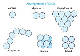
When we first came into class we handed in our lab reports if you finished them. They are do on Tuesday at 11:59 P.M. You can email it to Mr. Finley, but if your computer is not working Mr. Finley will not accept any excuses. Once the people who did their lab reports had them in, we started talking about how the bacteria reproduces. The equation you should use to find the amount of bacteria produced is two times whatever power you put in. For example, two to the second power equals four. Two to the third power equals eight and so on... We also came up with the statement, the more bacteria the faster they multiply. We call this binary fission. Binary fission means bacteria reproduces by splitting. Then, after all this we got our quizzes and graphs back. The grades were pretty good. The graph has the same pattern as the equation. Towards the end of the class, we started a mini project about different types of bacteria and there shapes. The different types were cocci, spirillum, diplo, strepto, and staphlyo. My group had cocci. We had to go on to smart notebook paste a picture and write a short description of what it looks like and the definition. Also, we had to describe how it is grouped. It should be short and just explain what needs to be explained. Tomorrow in class, we are suppose to present it to the class so they could learn what you learned


Stephen Ferraro

Period7

Thursday, October 28, 2010

Blog By Jake Vignali


Today in class Mr.Finley started off the period by talking about having the class observe their bacteria for the very last day. This is day seven, my group over the past week has had a-lot of bacteria growth. The most bacteria came from the middle school bathroom toilet. We also tested my locker which is 120A. We also tested the middle school bathrooms urinal. The last thing we tested the middle school bathroom mirror. The most bacteria growth came from the middle school bathroom toilet. there was a-lot of bacteria from others but the toilet had the most. Also we have mold covering nearly the entire peachery dish. The least amount of mold came from my locker corner. The percents of the mold growth is huge since the last time we checked out peachery dish. We had 52% of my locker covered in mold. There was 95% of the bathroom mirror covered. Then the toilet had 97% of bacteria covering it. Then finally we had the urinal which we found out had 80% covering it. I notice though that the locker corner had no bacteria at all. I think this is because the locker corner didn't come in contact with anything. So I came up with a hypothesis. It was that untouched areas will have less bacteria growth then contact areas. Like your foot would have less bacteria than your hand. Now we are going to observe bacteria that is macroscopic. Macroscopic is bacteria that is larger than microscopic bacteria but you still need a microscope to see it. But we are going to observe them in colonies (large numbers). And we are going to use a stereoscope to observe the colonies. A stereoscope is something you use to magnify things. Basically its something that you can see as a human but it helps you get a more detailed picture of the object like a spider. When I looked at the bacteria the mold was hairy. The bacteria had a bubble wrap feature to it. And that's how my day in science went. I have posted a picture though to show you the difference between bacteria and mold.
JV

Wednesday, October 27, 2010

Graphing Bacteria Growth


Today in class we discussed the graphs Period 7 did for homework last night. Some of the kids had questions. The graphs we did for homework were not that good so in class we went over how you could improve it. Some of the things students were missing in the graph was a trend line and titles. Also logic kids were missing. Some of the kids connected the dots and some didn't. Connecting the dots could help you figure out if you made a mistake or not. For the homework last night, we have to turn it in. We discussed how you could make it more presentable like not having a ripped piece of paper and you should use all of the space provided. We need to make sure that on the axis there is even spacing and also even intervals. On the y axis you had to make it up to 1 million. Then count and make sure its even. On the x axis you needed to have even spacing for the time. Also you need a key or a legend. For the homework you needed to have two separate lines representing the two different sets of data. For the different lines, you needed to represent them differently. Meaning different colored lines for the different sets of data on the graph. During the end of the period Mr. Finley let some of the kids redo there graphs because they did it wrong or they just didn't do it. For the kids that did it right with no errors are working on writing a conclusion for the graph. In the conclusion you should talk about the outcome of the experiment and any important things you noticed when you graphed the data. In the picture I attached in the top left corner shows trend in a graph.

Monday, October 25, 2010

Period 7

First, we did a warm-up problem with our tables. Second, we wrote down more observations and figured out how much more bacterial growth happened in our petri dishes. We wrote down more Qualitative data and found out what percent of each section has mold/bacteria growing in the petri dishes. Next, we played Trivial Pursuit because the online experiment didn't work.
RT

Friday, October 22, 2010

October 22nd P7 -JP

Period 7...By JP...10/22/10

First, we were talking about our lab reports which should have a hypothesis, conclusion, data, and your procedure. "For your procedure you must number your steps in order," mr. finley had stated. Just important reminders for your lab report. Second, Mr. finley talked about the observatioonal experimant that led to your hypothesis. You should probably include your observational experiment in your backround section. Your backround should be the first part of lab report. It sets up or makes a backround for your experiment. More specifically it is everything that leads to your experiment.(Important statements Mr. Finley made). Next we learned about our graghs. Basically Mr.Finley made sure we knew how to make a gragh.

Things to make a line gragh
1-Equal spaces between each axis lable.
2-Independent variable should be on the x axis.
3-Equal intervals between each axis lable.
4-Do not have to start at zero.
5-Scale should be an easy number to work with.
6-Lable each axis.
-Lable x axis with your independent variable.
-Lable your y axis gragh with your dependent variable.
7-Lables have to include units of measure.
8-Need a title
9-And ofcoarse put your data on your gragh.
10-Connect the points and/or draw the trend line.
11-Then try to find patterns or relationships between your independent variable and your dependent variable. This part is very important for understanding your gragh.
12-Your trend line does not have to be straight. Your trend line should touch some points and not touch others. A trend line is basically a genral line of the data on your gragh.

Next we worked on our graghs for our bacteria. If you missed class I can only say that everybody's bacteria grew. We did our usuall everyday basis of taking measurements, and adding to our graghs and coming up with more patterns.


email me at pricelax37@gmail.com,
or contact me on facebook for any questions.

Pic that may help you understand lab reports.


http://www.chem.fsu.edu/chemlab/chm1045lmanual/lab_report_table3.jpgThis is a table.

http://staff.argyll.epsb.ca/jreed/math9/strand4/graph_line.gif

That is a website leading to a picture that has a copyrite policy.


Thursday, October 21, 2010

Line Graphs

First we came to class. Then right away we got started on our experiment where we tested where bacteria is the most and the least. Also Mr. Finley told us to use the percent of the area in the Petri dish for amount of bacteria. We also took notes on the bacteria underneath the table we drew yesterday. The class had some very good questions like, "Are we doing the percent of the whole dish or just that section." The answer was the just the section. One group did a good job and got Bull Dog Bucks. They explained what they did. First they found out what it looks like out of the box and then divided it by the number of boxes. If any Table didn't finish went in during study hall. Then we started talking about line graphs. It usually shows trends or changes over time. Then he gave us an example on the white board. We had to make a line graph in our notebook to see if we knew how to make a line graph. Next we went over the independent and dependent variable for the graph. The dependent variable is the thing that is getting measured. The dependent variable goes on the y-axis. The independent variable always goes on the x-axis. Then on the smart board Mr. Finley drew a graph for the example on the white board.

-Johnny Shahpazian

Wednesday, October 20, 2010

Period 7

Yesterday in class we did an experiment which was to find a place where you think will contain bacteria, we took a cotton swab and rubbed it on a place where we thought there was bacteria. Then we rubbed the swab in the petri dish. 3/4 places we chose to test for bacteria were in the bathroom. We did this because we know tthe sections is because the places where we took the sample of aren't really clean places. hat bacteria is in places that aren't really clean and in dark places. I predict that bacteria will grow on all four sections of the pertri dish because we chose a bottom corner of a locker, the drain of a sink, a toilet handle, and a outer part of a urinal. The reason I think bacteria will grow in all of them is because I know that they aren't really clean places.

Today, web links were disabled I wwould've posted it but I couldn't anyway I found this article on lifewhile.com and it said the top 5 dirtiest places in school, bathroom was #3 on the list


- Jake Pickton

Tuesday, October 19, 2010

Experiment

We are talking about bacteria again. We are talking about where there is less bacteria, here are some places, the oven and a frezzer. There are more places but those are only some. We are also talking about where a lot of bacteria is, the air vents and underneath the wrestling mat and their are more. We are also making an experiment. We are going to make in a petry dish we are going to make 2 places where there is more bacteria and less bacteria. We took a sterilized swab rubed it agains our surface and then rubbed it on the part of the petry dish that has the initials of the surface we took. For example we took an air vent then rubbed it with the sterilized swab then rubbed it on the air vent corner. Now we can see tracks. We think the tracks are bacteria.


VP


Friday, October 15, 2010

Today we talked about conclusions and the result of our homeowork and our conclusions that we did. Mr.Finley showed us Ether Pad and how we can post pieces of evidence and most of the time we post our opinion. We reviewed the Lois Pasteur notes and simulation.On the notes Mr.Finley underlined each of the important notes. We talked about the Lois Pasteur simulation and experiment and how he tried to disprove the other peoples hypothesis'. I learned that mold is sitting in the air and people are very allergic to it. Mold basically just grows on dirty old objects or rotting food. In groups we discussed where in our school does mold and bacteria grow.

JPM